Building on these modular capabilities, we offer OEM-ready imaging solutions composed of combined high-performance capabilities from our advanced technology portfolio, purpose-built to support locally targeted bronchoscopic and surgical interventions across key therapeutic areas.
These solutions are designed for integration into clinical decision-support environments deliver quantitative, anatomically precise insights to support:
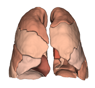
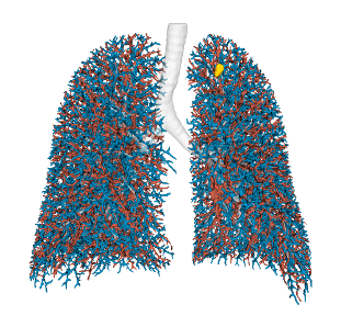
- Treatment target identification
- Patient selection and eligibility assessment
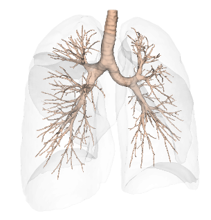
- Pre-operative planning of virtual bronchoscopy pathways
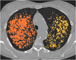
- Post-treatment evaluation and response monitoring
Developed and validated on disease-specific datasets, our solutions deliver reliable performance and lasting value, continuously refined through research and clinical collaboration.
Our software solutions are ready to be applied to a range of targeted treatment approaches, such as: